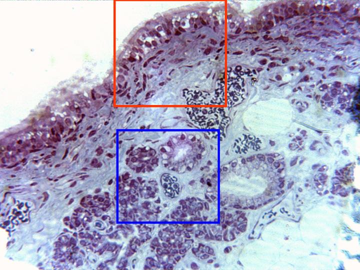
Semifine cross-section of trachea stained with toluidine
blue and observed with the objective of 40x. This image
shows the different layers that constitute the trachea:
mucous,
constituted by pseudostratified prismatic epithelium,
submucous,
formed by connective tissue containing abundant
blood vessels,
and
glands.
Details of the mucous and the glands are observed in
other microphotographies (zones framed in red and blue
respectively)