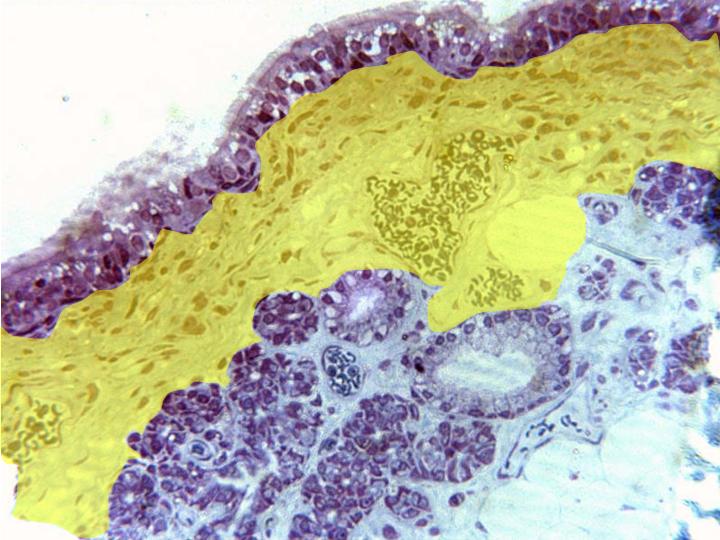
Semifine cross-section of trachea stained with toluidine
blue and observed with the objective of 40x. This image
shows the different layers that constitute the trachea:
mucous,
constituted by pseudostratified prismatic epithelium,
submucous (yellow),
formed by connective tissue containing abundant
blood vessels,
and
glands.